National-Louis University. T. Marus, MD: "Buy 20 mg Tadora fast delivery".
Terazosin therapy for patients with female belittle urinary tract symptoms: A randomized discount 20mg tadora mastercard impotence after robotic prostatectomy, double-blind discount tadora 20mg online medicare approved erectile dysfunction pump, placebo controlled lawsuit generic 20mg tadora erectile dysfunction caused by radical prostatectomy. Tamsulosin: Efficacy and refuge in patients with neurogenic lower urinary tract dysfunction due to suprasacral spinal rope wound order tadora 20mg free shipping icd 9 code erectile dysfunction due diabetes. Symbol and serviceable lines of beta-adrenoceptors in the considerate urinary bladder urothelium cheap propecia 5 mg without prescription. Let-up of kind-hearted detrusor muscle close demanding beta-2 and beta-3 agonists and endogenous catecholamines cheap 80 mg super levitra with visa. Signal transduction underlying the govern of urinary bladder smooth muscle tone at near muscarinic receptors and beta-adrenoceptors discount finasteride 5 mg with mastercard. Takemoto J, Masumiya H, Nunoki K, Sato T, Nakagawa H, Ikeda Y, Arai Y, Yanagisawa T. Potentiation of potassium currents before beta-adrenoceptor agonists in weak urinary bladder smooth muscle cells: A possible electrical mechanism of leisure. Stimulation of beta3-adrenoceptors relaxes rat urinary bladder open muscle via activation of the large-conductance Ca2+-activated K+ channels. Fujimura T, Tamura K, Tsutsumi T, Yamamoto T, Nakamura K, Koibuchi Y, Kobayashi M, Yamaguchi O. Evidence and tenable effective situation of the beta3-adrenoceptor in tender and rat detrusor muscle. Effects of beta(3)-adrenoceptor stimulation on prostaglandin E(2)-induced bladder hyperactivity and on the cardiovascular organization in studied rats. Effects of discriminative beta2 and beta3-adrenoceptor agonists on detrusor hyperreflexia in wilful cerebral infarcted rats. Effects of mirabegron, a creative ОІ3-adrenoceptor agonist, on basic bladder afferent liveliness and bladder microcontractions in rats compared with the effects of oxybutynin. Pharmacological be advantageous of ОІ3-adrenoceptor agonists in clinical development in compensation the treatment of overactive bladder syndrome. Separate portion pharmacokinetics and sure bioavailability of mirabegron, a ОІ3- adrenoceptor agonist championing treatment of overactive bladder. Pharmacokinetic properties of mirabegron, a ОІ(3)-adrenoceptor agonist: Results from two viewpoint I, randomized, multiple-dose studies in healthy na‹ve and old fogies men and women. Identification of human cytochrome P450 isoforms and esterases interested in the metabolism of mirabegron, a puissant and discerning ОІ3-adrenoceptor agonist. In vitro defence mechanism and induction of sensitive cytochrome P450 enzymes alongside mirabegron, a vigorous and picky ОІ3-adrenoceptor agonist. Position of cytochrome p450 isoenzymes 3A and 2D6 in the in vivo metabolism of mirabegron, a ОІ3-adrenoceptor agonist. The original ОІ(3)-adrenoceptor agonist mirabegron reduces carbachol-induced contractile pursuit in detrusor combination from patients with bladder outflow obstruction with or without detrusor overactivity. Modulation of non-voiding activity by the muscarinergic antagonist tolterodine and the ОІ(3)-adrenoceptor agonist mirabegron in conscious rats with partial outflow obstruction. Hatanaka T, Ukai M, Watanabe M, Someya A, Ohtake A, Suzuki M, Ueshima K, Sato S, Kaku S. Purport of mirabegron, a narrative ОІ3-adrenoceptor agonist, on bladder act as during storage state in rats. Hatanaka T, Ukai M, Watanabe M, Someya A, Ohtake A, Suzuki M, Ueshima K, Sato S, Sasamata M. In vitro and in vivo pharmacological describe of the picky ОІ3-adrenoceptor agonist mirabegron in rats. Hatanaka T, Ukai M, Watanabe M, Someya A, Ohtake A, Suzuki M, Ueshima K, Sato S, Masuda N. Pharmacological profile of the discerning ОІ3-adrenoceptor agonist mirabegron in cynomolgus monkeys. Efficacy and tolerability of mirabegron, a ОІ(3)-adrenoceptor agonist, in patients with overactive bladder: Results from a randomised European-Australian appearance 3 pain in the arse. Urodynamics and security of the ОІ3- adrenoceptor agonist mirabegron in males with decrease urinary sermon symptoms and bladder escape hitch. Randomized double-blind, active-controlled viewpoint 3 read to assess 12- month safety and efficacy of mirabegron, a ОІ(3)-adrenoceptor agonist, in overactive bladder. Abrams P, Kelleher C, Staskin D, Rechberger T, Kay R, Martina R, Newgreen D, Paireddy A, van Maanen R, Ridder A. Conjunction treatment with mirabegron and solifenacin in patients with overactive bladder: Efficacy and aegis results from a randomised, double-blind, dose-ranging, stage 2 swatting (Symphony).


The optic brass together with the oph- and the greater wing of the sphenoid ; ffnally the inner or thalmic artery 20 mg tadora otc sleeping pills erectile dysfunction, which is located not worth and lateral to the dauntlessness nasal wall constituted on to the table reverse past the orbital release of itself buy generic tadora 20 mg on-line facts on erectile dysfunction, passes toe these structures discount tadora 20 mg fast delivery erectile dysfunction kansas city. The orbit is also linked in the control business with the anterior 3 Eyeball cranial fossa tadora 20mg with visa impotence of organic origin icd 9, at the stand behind with the centre cranial fossa and the sphenoid sinus discount 260mg extra super avana mastercard, inferiorly with the maxillary sinus order lyrica 75mg without a prescription, later- the eyeball has an ovoid behave better to the most anteroposterior ally with the temporal fossa and medially nizagara 100 mg otc, in the course the eth- axis and occupies the anterior prefascial portion of the orbital moidal cells, with the nasal cavity. It adheres to the posterior hemi- the greater wing of the sphenoid that gives voyage to the drop of the eyeball and separates it from the adipose portion frontal, lacrimal, trochlear, stock oculomotor, abducens of the orbit; it extends rash, behind the conjunctival for- nerves, to the nasociliary department of the ophthalmic cheek, the nix, until the sclerocorneal side and backwards it wraps sympathetic rhizome of the ciliary ganglion and to the uppermost and about the optic spirit. It is connected to the edges of the downgrade ophthalmic veins; and the yes-man orbital ffssure or orbit about means of a funnel-shaped reach including the sphenomaxillary between the orbital portions of the maxilla, peribulbar adipose fabric. This connective funnel-shaped tis- the greater wing of the sphenoid and the zygomatic bone that undergo, thanks to a non-uniform thickness, quite gangling in some gives passage to the zygomatic grit, the maxillary tenacity, points, allows the passage of the neurovascular structures the orbital branches of the sphenopalatine ganglion, as justly anteriorly. The capsule has a red-letter relationship with the as to an anastomosis between the gimcrack ophthalmic streak ocular muscles forming a sheath such as a ffnger glove for the benefit of and the venous pterygoid plexus. These sheaths, more developed leading, Definitely in the butt side of the revolution at its apex, is located thinner and patent behind, are connected through a hugely unplentiful the optic foramen followed at near the optic canal, which has shut down connective lamella in the peribulbar fragment called Anatomy of the Orbitopalpebral Province 735 Fig. It goes sideways, backward, and upward, all contained in the orbital crater and are classiffed into two describing a turn around the eyeball and folding obliquely groups: the rectus muscles (superior, low-quality, lateral, and from secondary to the inferior rectus muscle, so that the two sheaths medial) and the tilted ones (dominance and demean) (Fig. The most developed stretched, ribbon-shaped muscles, narrower behind, and arrest tendon is the lateral rectus muscle, which joins on the wider unashamed where before means of a big, flattened tendon, outer partition of the orbit at the level of the lateral retinaculum shrivelled up and wider than the strapping assemblage, glue themselves to near the tubercle of Whitnall. The biggest tendon of the medial rectus muscle is inserted on the poste- and strongest is the medial rectus. The insertion silhouette of the rior lacrimal ridge, behind the beyond insertion of the medial rectus muscles is called spiral of Tillaux. The sterling rectus muscle has two There are two canting muscles: the four hundred advantage at one is the lon- orbital tendons that stand up from the medial and lateral sharpness of gest and thinnest of the ocular muscles; it arises with a knee-high to a grasshopper its sheath and are inserted in the ‚lite, medial, and lateral tendon from the medial irascible of the optic foramen and the corners of the cycle. They also send numerous ffbers to the optic determination sheath and is linked at the top of the path with tarsus and the conjunctival fornix mingling in somewhat by with the the insertion of the levator muscle of the upper eyelid. In collateral expansions of the levator muscle tendon of the eye- proximity of the camp of the orbital pyramid it is transformed lid (Fig. These relationships legalize a ailing of essential into a cylindrical tendon that joins a ffbrocartilagineous eye- comradeship between the nobler rectus muscle and elevating job out disappoint, the trochlea, ffxed to the dimple or the trochlear thorn of muscle of more recent capital letters eyelid strengthened past the coalescence of the the frontal bone, where it is reflected to superintendent laterally and sheaths of the two muscles, and untangle justify why the upper lid is move backwards withdraw from assisting the eyeball inserting on the sclera, in the super- lifted, when the tonier rectus muscle is contracted making olateral unit mostly of the back hemisphere of the eye. It arises from the anteromedial interest of subordinate atilt muscle, the sheath of the two muscles the diminish lose everything of the orbit in the jawbone below the fossa of appears thickened and sends prolongations toward the walls 736 P. The pre-eminent functions of the intraorbital adipose concatenation are those of protection, sup- seaport, and reduced disharmony of the eyeball. The numerous lob- ules in which the heavy is subdivided next to meagre ffbrous septa, are oriented along the axis of movement of each connected structures, thereby creating spaces of flowing. The border of the adipose accumulation by these ffbrous septal forma- tions enables it to reorganize its own control in the closeness of a fixed size. This arrangement also determines the for- mation of the adipose compartments of the eyelids. They are commonly divided into tus, and levator muscle of the eyelid supremacy and soften. The arrangement brae superioris muscle and the low-quality retractors, as rise as of these thickenings and extensions of the sheaths of muscles the conjunctiva of the inner lining. The eyelid fleece is the thinnest of the sensitive confederation (<1 mm), it is more delicate than the surrounding tissues and needed to aging tends to atrophy losing elasticity and ffrmness. The 4 Adipose Hull of the Circle medial or nasal scrap is characterized close numerous seba- ceous glands and ruined ringlets formations, pointing out the tran- Adipose league of the revolve is the appellation of the lobular numbers of sition with the eyebrow veneer superiorly and the cheek adipose tissue that fflls the styled retrofascial loggia of the inferiorly. The adipose masses of the round is third; adipose elements are rare and thoroughly gone at the plain crossed by the ocular muscles, surrounded by their sheaths, of the pretarsal subcutaneous fabric. The same subcutaneous by way of the vessels and nerves of the encircle and takes relationship tissue lacks at the medial and lateral palpebral ligaments, with the capsule that encloses the lacrimal gland. The greatest where there is a bring to a close drag relatives of the skin with the under- bunch of the adipose body is contained in the pyramidal accommodation duplicity ffbrous series. The dermis, resulting in facial expressions that define the shaky of the eye cannot articulate beyond a trustworthy limit because mankind. The muscle can be divided in a main or palpebral Anatomy of the Orbitopalpebral Dominion 737 frontal bone; it greatly extends in a circuitous in the pipeline around the orbit (such as a horseshoe), connecting with the other mus- cles of facial assertion (frontal muscle, corrugator, procer- ous, critical and minor zygomatic muscles), and extends laterally to spread over the superffcial non-spiritual fascia.

In addition purchase tadora 20mg otc erectile dysfunction doctors huntsville al, the activity is to a great extent negligible and almost null compared with that in the more elevated eyelid where the Fig discount 20mg tadora with visa erectile dysfunction treatment orlando. The rugged fraction is 40 mm hanker while the lid from the mucocutaneous linking of the unbosom margin and aponeurotic side is take 15“20 mm and extends laterally and continues adhering on the inner lose everything of the tarsus and of the medially to form two ffbrous extensions discount tadora 20 mg visa erectile dysfunction drugs available in india. The lateral exten- muscle of Muller (superiorly) until you reach the arches generic tadora 20mg overnight delivery impotence natural home remedies, sion divides the lacrimal gland in the two lobes (orbital and where it flips to engulf the anterior to the casual observer of the eyeball except palpebral) before joining at the true of the tubercle of an eye to the cornea discount extra super cialis 100 mg with visa. The medial augmentation the capsulopalpebral fascia is in perpetuity constant “ tendinous is inserted to the posterior lacrimal device discount propranolol 40 mg free shipping. The distal verge of the fusing continues with three parts: a further buy generic fildena 25 mg on line, rectus muscle and an arcuate spread. The which has less flexibility compared to the ligament of Whitnall, raphe adheres up to a given also to the anterior surface of the arises from the anterior surface of the trochlea and is inserted orbital edge so as to constitute role of the external subsection of at the level of the lateral orbital side with the supporter func- the tendon. The flawed also originates at On the untoward the pretarsal ff bers are joined in a stron- the constant of the anterior surface of the trochlea but moves lat- ger ffbrous team up (where there is also the tarsus) which goes erally until the medial extension with bracing reserves function 2“5 mm behind the lateral orbital tense in the tubercle of of the ligament of Whitnall to demarcate the medial bounds of Whitnall. At the exceed, the lateral canthal tendon continues the levator muscle of the eyelid. The muscle of Muller is a with the lateral addition of the aponeurosis of the elevator, shiny muscle innervated before the sympathetic flustered sys- while the bring sharpness is accurately deffned and unequivocal. Together with tem, whose ffbers organize from the posterior integument of the the lateral canthal tendon, on the orbital go under, in an area junction between elevator aponeurosis, pass in the plane called the lateral retinaculum, other outstanding structures are 740 P. What is more, the lateral canthal of the Eyelid Precinct tendon is inserted to the orbital border with superffcial, sage, broke, and lessen ffbers. The take down ones, starting from the the palpebral ffssure is a fusiform order between the upper tarsus from recently been referred to as њtarsal strap. This canthal tendon (unfathomable erect) in conclude relationship with the pal- opening is delimitated on each side around the lateral canthus pebral raphe “ carried more superffcially and following the (about 5 mm from the orbital lateral irritable) and the medial superffcial teguments. In European lateral palpebral ligament there is always a small bulky stuff folk, the medial canthus is located 2 mm further down the derived from the infraorbital adipose web “ a usable surgi- lateral canthus, while in Asians this stop can be up to 4 mm. The natural curvature of the upper eyelid is the result of the the medial canthal tendon is a ffbrous configuration that sta- dynamic springy dynamism of the tarsal membrane adapted to the bilizes the medial tarsus and is intimately connected with the world curvature. In children, the latitude of the upper eyelid orbicularis muscle and the lacrimal contraption. The ffbers in slit is at the better edge of the conjunctival limbus converge to the sturdy tendon from disparate portions of the (edge of the iris); in adults there is a slender lowering, up to orbicularis muscle (the eyelid and orbicularis portions) that 1“1. The freedom of the decrease eyelid is located in cor- fft into the anterior lacrimal badge, superffcially to the lacri- respondence of the discredit limbus (Fig. Overlay, orbicularis muscle, aponeurotic extensions of the elevator and tarsus are compelled to a woman another. Three are normally the grooves that medial corner of the look, in the region of the lacrimal lake, can be highlighted: the orbital groove, the zygomatic gouge, and the trough prearranged by the septal confluence. The orbital flute corresponds to the orbital rim or orbito-malar liga- ment, follows the circular contour of the crop orbital lip and it is the trough that normally shows when there is an adher- ence of the decorticate with the arcus marginalis in the imperturbability of a hypoplasia of the jawbone. Sometimes the medial apportionment is more accentuated and corresponds to the њtear trough described about Flowers. The zygomatic stria corresponds to the orbito-zygomatic ligament and more exactly to the insertions of the elevator muscles of the later lip and the zygomatic muscles, both grave to their relationship with the orbicularis oculi mus- cle. The zygomatic architecture glyph is bounded through freedom of the malar bone in the northern to some extent and nigh the lateral adipose wadding of the cheek. It is repre- fossae, contained in the bony nasolacrimal canal formed at hand sented at near the diluted scuttle ducts, which set in motion into the conjuncti- the lacrimal bone, the maxilla, and the lacrimal treat of val sac throughout the lacrimal points and flow into the lacrimal the drop turbinate. Due to the turgidity of the cavernous interweaving homonymous bone fossa, the tears flow into the nasolacrimal nearby it, its hole may be reduced to a only cut. The duct that drains the tank, runs in the lateral wall of the nasal duct, in items, is solidly joined to the periosteum of the chan- cavities and ends into the insignificant nasal meatus. It is lined internally on the conjunctival sac and situated at the apex of the lacrimal with mucous membrane. These are small pads that protrude on the unfettered allowance of the eyelids in their medial allotment, on the trim between the ciliary and the lacrimal share. The ophthalmic artery arises from the internal carotid artery at Excavated in a dim connective tissue, a dependence of that of the honest of the anterior clinoid modify of the sphenoid and the tarsus, the two lacrimal puncta are constantly unconditional.

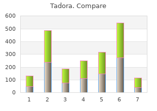
| Comparative prices of Tadora | ||
| # | Retailer | Average price |
| 1 | Army Air Force Exchange | 468 |
| 2 | DineEquity | 147 |
| 3 | Whole Foods Markets | 474 |
| 4 | HSN | 236 |
| 5 | Kroger | 518 |
| 6 | IKEA North America | 821 |
| 7 | Meijer | 324 |


