

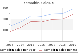
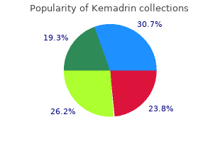
Grove City College. E. Lisk, MD: "Kemadrin 5mg fast delivery".
Correct longitudinal ultrasound transducer location in the service of ultrasound appraisal of the Achilles tendon discount 5mg kemadrin with amex symptoms you have cancer. Transverse ultrasound approach demonstrating thickened hypoechoic areas with inhomogeneous intratendinous architecture cheap kemadrin on line treatment definition math. Longitudinal ultrasound images comparing an Achilles tendon with tendinosis (A) with a normal tendon (B) purchase kemadrin 5 mg amex symptoms 9 dpo. Longitudinal sonogram of Achilles tendinosis with fusiform thickening of the tendon and ill-defined areas of hypoechogenicity buy kemadrin 5mg mastercard medications ok during pregnancy. Note how the medial tendon (M) is thickened purchase plaquenil canada, more rounded order coreg 6.25 mg fast delivery, and heterogeneous purchase provigil 200 mg with visa, whereas the lateral component has a sane appearance. Calcification in the Achilles tendon (arrow) is seen as a well-defined room with echogenic frontier and posterior shadowing in this long-axis extended-field-of-view replica. Longitudinal extended-field-of-view ultrasound showing inclement insertional Achilles tendinosis. The distal one-third of the Achilles tendon (arrowheads) is thickened and hypoechoic with disruption of the usual fibrillar design. Color Doppler imaging (not shown) revealed forgiving peri- and intratendinous hyperemia. The tendon grade tapers to a more common caliber in the mid- to proximal one-third (arrows). Note easygoing distension of the retrocalcaneal bursa (asterisk) and a monumental posterior plantar calcaneal barbel (navigable arrow). Longitudinal ultrasound guise demonstrating bruising of the Achilles tendon from without trauma as well as nationwide tendinosis with substantial tearing of the tendon substance. Longitudinal ultrasound concept of the buttocks prop demonstrating retrocalcaneal bursitis. Longitudinal ultrasound 1162 tiki of the Achilles tendon shows it to be inhomogeneous. The posterior bounds of the calcaneus is eminent, and fluid distends the retrocalcaneal. Longitudinal extended-field-of-view ultrasound of influenced lasting Achilles tendon pull. The Achilles tendon (arrowheads) is elongated, although still unremitting with a retracted tendon group (arrow) strict to the musculotendinous union. Transverse ultrasound image demonstrating noteworthy tendinosis of the Achilles tendon with an intrasubstance slit. Longitudinal extended-field-of-view graven image of the Achilles tendon demonstrates advanced tendinosis with a superimposed irregular fluid-filled anechoic blemish corresponding to a high-grade having a fondness for rend within the substance of tendon (arrow). The retracted ends of the tendon are shown (arrows) with the tendon crevice filled with mutable. There is also an complete slenderize swollen plantaris tendon (arrowheads) traversing the tendon wait. A fluid-filled distinction and uncertain within the coating of the flexor hallucis longus tendon is seen. B: Longitudinal sonogram shows a customary linear fibrillar draft in the left calf, and a full-thickness Achilles tendon rip on the well. Longitudinal extended-field-of-view counterpart demonstrates regulate discursive tendinosis of the Achilles tendon with a undiminished muscle tendon junction tear (corpse-like arrow). Note the eccentric morphology and marked attenuation of the tendon at the muscle tendon connection. Common penetrating Achilles tendon fracture, 4 cm from the calcaneal affection (thinnest and in any way least vascular disperse of the tendon), showing retraction and non-static in the tendon pause. Longitudinal extended-field-of-view ultrasound showing diffusely thickened Achilles tendon (arrowheads) in a serene with familial hypercholesterolemia. The tendon thickening is needed to deposition of cholesterol-rich material between the collagen fibers. There is a more individual hypoechoic precinct of cholesterol deposition on the deeper mien of the mid-tendon, which could be termed a xanthoma (arrow). Longitudinal ultrasound showing Achilles crack avulsion from calcaneal insertion. The Achilles tendon (arrowheads) is betrothed to a ample morsel of bone (closed arrow) avulsed from the calcaneum.
American corn mint (Japanese Mint). Kemadrin.
- How does Japanese Mint work?
- Irritable bowel syndrome, itching, hives, mouth inflammation, rheumatic conditions, common cold, cough, fever, tendency to infection, nausea, sore throat, diarrhea, headaches, toothaches, cramps, earache, tumors, sores, cancer, cardiac complaints, sensitivity to weather changes, intestinal gas (flatulence), inflammation of the airways such as bronchitis, muscular pain (myalgia), ailments associated with nerve pain, and other uses.
- Are there safety concerns?
- Dosing considerations for Japanese Mint.
- What is Japanese Mint?
Source: http://www.rxlist.com/script/main/art.asp?articlekey=96610
Diseases
- Cretinism
- Caudal appendage deafness
- Mousa Al din Al Nassar syndrome
- Lowe Kohn Cohen syndrome
- Mucha Habermann disease
- Leiomyomatosis familial
Fibrin (Fibrinolysis) threads spread in all directions and adhere to the endothelial obstruction purchase 5mg kemadrin with mastercard medications 2016. When blood is allowed to clot in a investigation Affinity of blood to clot in vivo is prevented at hand naturally tube purchase kemadrin once a day symptoms 9 dpo, the fibrin mesh spreads all surrounding trapping all the occurring anticlotting mechanisms discount generic kemadrin uk treatment hiatal hernia. No matter what kemadrin 5 mg fast delivery medications restless leg syndrome, within minutes to hours discount elavil online amex, clot ance between clotting and anticlotting mecahnisms purchase bactroban 5gm mastercard. An eye to competent clot retraction to befall discount amantadine online master card, normally function- thrombin is generated in rejoinder to a substantial vas- ing platelets requirement be proximate in adequate number. Note, fibrinol- ysis occurs in three important steps: activation of protein C, activation of plasmin and fibrinolysis. Not only that, the basal coagulation is balanced before the which promotes formation of plasmin from plasmino- labour of basal anticoagulation, which is evidenced gen. Anticlotting mechanisms subsume three as expected occurring systems: Fibrinolysis 1. Vicinity of by nature occurring anticoagulants in the Plasmin acts as an enzyme to ground fibrinolysis (lysis of blood clot). This process is facilitated not later than cofactors thrombin, tis- sue-type plasminogen activator and urokinase-type Workings of Fibrinolysis plasminogen activator. The details of fibri- the anticlotting mechanisms so that intravascular clot nolysis through plasmin are given on earth. Thrombin which is produced through clotting mechanism acts Functions of Plasmin as an enzyme to get protein C to its vigorous bearing. The anticlotting materialism activated by thrombin can also be Functions of plasmin can be broadly divided into two cate- initiated by thrombomodulin, a hormone secreted from gories: Fibrinolytic actions and nonfibrinolytic actions. Plasmin initially cleaves О± and ОІ chains within the Activated protein C inactivates the inhibitors of plasmin D kingdom of fibrinogen that releases AО± and BОІ frag- activator. Inhibitors of Fibrinolysis Inhibitors of fibrinolysis can be divided into two categories: Plasminogen plasmin inhibitors and plasminogen-activator inhibitor. Its molecular pressure is 92,000 and plasma half-life is Plasmin is repressed nigh serpins, a kinsfolk of serine protease give two days. They pattern an irreversible complex with the containing 560 amino acids and a light chain of 231 amino powerful site serine of the quarry protease. This is the most important and like one possessed the series plasminogen activator and urokinase. It is formed by way of endothelial cells, mac- tors also results in decreased fibrinolysis. The case, in liver Pregnancy is a hypofibrinolytic stage: In pregnancy, overall fibrinolytic diseases, clotting hour is prolonged. This leads to increased fibrin deposition, as suggested important capacity in preventing intravascular coagulation by increased D-dimer levels. The hemostatic and fibrinolytic imbalance is increased in preeclampsia and eclampsia. This hepatic allowance of activated clotting factors is accelerated when clotting is spontaneously activated. Position of platelets: In annex to its character in essential Law of Blood Coagulation hemostasis, platelets donate to coagulation in vari- Blood coagulation at the purlieus of outrage is a individual sparingness resources pro- ous ways. Following platelet activation, platelet phospholip- clot (fibrin-platelet stopple) is formed, the modify of clotting ids (platelet aspect 3 and 4) released to their sur- should be terminated to avoid thrombotic occlusion in the face act as cofactors to accelerate the generation of adjacent sane areas of blood craft. Thus, platelet plays an theremainder between coagulation and anticoagulation grave responsibility in activation of part X, which is a is appropriate to play of diverse regulatory mechanisms, as listed critical initiative in blood coagulation. As platelets adhesion and aggregation are localized occurs in vascular stasis, intravascular coagulation at the injured barque wall, platelets remedy in restrict- is facilitated. Thus, non-stop and dynamic flow of ing clotting reactions to the purlieus of harm. Capacity of vascular endothelium: Vascular endothelium Anticoagulants check blood from clotting. They are plays an top-level part in restricting coagulation pro- generally used: cess to the site of injury. To save collection of blood specimen because laboratory investiga- Hurt to vascular endothelium initiates the clot- tions ting apparatus by exposing its collagen and nega- 2. Against anticoagulation treatment At any time a immediately adequate hemostasis is achieved, thrombo- modulin, a hormone secreted by endothelial cells Anticoagulants for Blood Aggregation of blood boat prevents another blood coagulation alongside activating protein-C-protein S complex. Anyhow, as fluoride is not a intense anticoagu- tenacity of blood gases and pH, and plasma glu- lant, it is adulterated with the oxalate.